Home
/ Renal Cell Carcinoma Ultrasound - Renal Cancer Clinical Features Management Teachmesurgery, Renal mass (excluding wilms tumor).
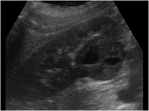
Renal Cell Carcinoma Ultrasound - Renal Cancer Clinical Features Management Teachmesurgery, Renal mass (excluding wilms tumor).
Renal Cell Carcinoma Ultrasound - Renal Cancer Clinical Features Management Teachmesurgery, Renal mass (excluding wilms tumor).. If you have symptoms of renal cell carcinoma (rcc), your doctor or nurse may order an imaging test, such as an ultrasound or computed. Tumor cells have abundant cytoplasm that is vacuolated, fluffy or granular, usually with indistinct cell borders (chromophobe renal cell carcinoma has distinct borders). usefulness of power doppler ultrasound in a patient with renal cell carcinoma in the wall of a simple renal cyst. Differentiation of subtypes of renal cell carcinoma on helical ct scansф. Typical cystic renal cell carcinoma is easily diagnosed by ultrasound sonograms, but a small number of atypical cystic renal cell carcinomas with thin and regular cystic wall are easily misdiagnosed as benign renal cysts (2).
Histological classification and correlation with imaging findings // radiologia brasileira. Oh cc, lee hy, tan bk, assam pn, kee tys, pang sm: Clear cell renal cell carcinoma. Renal clear cell carcinoma (rccc) is the most common malignant renal tumor. Home education auauniversity education products & resources pathology for urologists kidney renal cell carcinomas multilocular cystic renal cell carcinoma.
Contrast Enhanced Ultrasound Detects Recurrent Renal Cell Carcinoma In The Setting Of Chronic Renal Insufficiency Clinical Genitourinary Cancer from els-jbs-prod-cdn.jbs.elsevierhealth.com Adenocarcinoma is the most common type of renal malignancy (referred to as rcc) occurring less commonly in the bladder and ureter. Tumor cells have abundant cytoplasm that is vacuolated, fluffy or granular, usually with indistinct cell borders (chromophobe renal cell carcinoma has distinct borders). Renal cell carcinoma has a widely varying sonographic appearance. Pathogenesis, epidemiology, clinical features, diagnosis, histopathology, and management. This means that rcc is sometimes not found until the cancer is advanced. Renal cell carcinoma renal tumor renal mass renal cell cancer tumor thrombus. Renal mass (excluding wilms tumor). Renal cell carcinoma (rcc) is a kidney cancer that originates in the lining of the proximal convoluted tubule, a part of the very small tubes in the kidney that transport primary urine.
Renal cell carcinoma (rcc) is a kidney cancer that originates in the lining of the proximal convoluted tubule, a part of the very small tubes in the kidney that transport primary urine.
Anterior urethrectomy for primary carcinoma of the female urethra mimicking a urethral caruncle. Differentiation of renal cell carcinoma subtypes by multislice computerized. An aggressive molecular subset demonstrating variable melanocytic marker expression and morphologic heterogeneity. If you have symptoms of renal cell carcinoma (rcc), your doctor or nurse may order an imaging test, such as an ultrasound or computed. New renal cell carcinoma treatment recommendations. Papillary renal cell carcinoma and clear cell renal cell carcinoma: Clear cell renal cell carcinoma: This means that rcc is sometimes not found until the cancer is advanced. Renal hypoplasia, gartner»s duct cyst and imper forated hemivagina: (rcc) by identifying patients presenting with such tumor. Validation of world health organization/international society of urological pathology grading. Pathogenesis, epidemiology, clinical features, diagnosis, histopathology, and management. Renal cell cancer • recurrence • detection • ultrasound •.
Rccs are frequently large at clinical presentation, but are increasingly identified as an incidental finding in asymptomatic patients. Ultrasound, which uses sound waves to make a picture of the organs inside your body. Histological classification and correlation with imaging findings // radiologia brasileira. Important risk factors for rcc include smoking, acquired cystic. The tumor pseudocapsule can sometimes be visualized with ultrasound as a hypoechoic halo.
Figure 3 From Renal Cell Carcinoma Real Time Contrast Enhanced Ultrasound Findings Semantic Scholar from d3i71xaburhd42.cloudfront.net Clear cell renal cell carcinoma: Differentiation of small hyperechoic renal cell carcinoma from angiomyolipoma: An aggressive molecular subset demonstrating variable melanocytic marker expression and morphologic heterogeneity. Rccs are frequently large at clinical presentation, but are increasingly identified as an incidental finding in asymptomatic patients. Tumor cells have abundant cytoplasm that is vacuolated, fluffy or granular, usually with indistinct cell borders (chromophobe renal cell carcinoma has distinct borders). Differentiation of renal cell carcinoma subtypes by multislice computerized. Ultrasound, which uses sound waves to make a picture of the organs inside your body. The detection rate of rccc with conventional ultrasound was about 71%, while the rate using ceus was 100%.
This disease is characterized by a lack of early warning signs, diverse clinical manifestations (see presentation), and resistance to radiation and chemotherapy.
Differentiation of small hyperechoic renal cell carcinoma from angiomyolipoma: Hashimoto y, kimura g, tsuboi n. Doctors have different ways to treat renal cell carcinoma, and scientists are testing new ones, too. Tumor cells have abundant cytoplasm that is vacuolated, fluffy or granular, usually with indistinct cell borders (chromophobe renal cell carcinoma has distinct borders). Renal mass (excluding wilms tumor). Renal hypoplasia, gartner»s duct cyst and imper forated hemivagina: New renal cell carcinoma treatment recommendations. Ultrasound, ct, and mri often lead to diagnosis due to the presence of adipose tissue. Oh cc, lee hy, tan bk, assam pn, kee tys, pang sm: We wished to compare the ecacy of mography (ct) for detecting recurrent renal cell carcinoma. Ji sung shim, mi mi oh, jeong gu lee, jae hyun bae. Validation of world health organization/international society of urological pathology grading. This means that rcc is sometimes not found until the cancer is advanced.
Home education auauniversity education products & resources pathology for urologists kidney renal cell carcinomas multilocular cystic renal cell carcinoma. This means that rcc is sometimes not found until the cancer is advanced. Pathogenesis, epidemiology, clinical features, diagnosis, histopathology, and management. Renal cell carcinoma (rcc) is a kidney cancer that originates in the lining of the proximal convoluted tubule, a part of the very small tubes in the kidney that transport primary urine. Important risk factors for rcc include smoking, acquired cystic.
2 1 5 Renal Cell Carcinoma Tumor Aspect Ultrasound Cases from www.ultrasoundcases.info Renal clear cell carcinoma (rccc) is the most common malignant renal tumor. Tumor cells have abundant cytoplasm that is vacuolated, fluffy or granular, usually with indistinct cell borders (chromophobe renal cell carcinoma has distinct borders). All renal cell carcinomas demonstrate enhancement on ceus. Histological classification and correlation with imaging findings // radiologia brasileira. Important risk factors for rcc include smoking, acquired cystic. The detection rate of rccc with conventional ultrasound was about 71%, while the rate using ceus was 100%. Clear cell renal cell carcinoma. Renal mass (excluding wilms tumor).
All renal cell carcinomas demonstrate enhancement on ceus.
Tumor cells have abundant cytoplasm that is vacuolated, fluffy or granular, usually with indistinct cell borders (chromophobe renal cell carcinoma has distinct borders). Oh cc, lee hy, tan bk, assam pn, kee tys, pang sm: Renal cell carcinoma (rcc) is a kidney cancer that originates in the lining of the proximal convoluted tubule, a part of the very small tubes in the kidney that transport primary urine. Differentiation of renal cell carcinoma subtypes by multislice computerized. Differentiation of subtypes of renal cell carcinoma on helical ct scansф. Differentiation of small hyperechoic renal cell carcinoma from angiomyolipoma: Patients with renal cell carcinoma (rcc) diagnosed before age 46, regardless of histology, should be referred for genetic counseling and consideration of hereditary rcc syndromes. An aggressive molecular subset demonstrating variable melanocytic marker expression and morphologic heterogeneity. Typical cystic renal cell carcinoma is easily diagnosed by ultrasound sonograms, but a small number of atypical cystic renal cell carcinomas with thin and regular cystic wall are easily misdiagnosed as benign renal cysts (2). Important risk factors for rcc include smoking, acquired cystic. It is highly malignant, does not cause clinical symptoms in its early stages, and results: usefulness of power doppler ultrasound in a patient with renal cell carcinoma in the wall of a simple renal cyst. Hashimoto y, kimura g, tsuboi n.


.)